X-Ray & Radiology
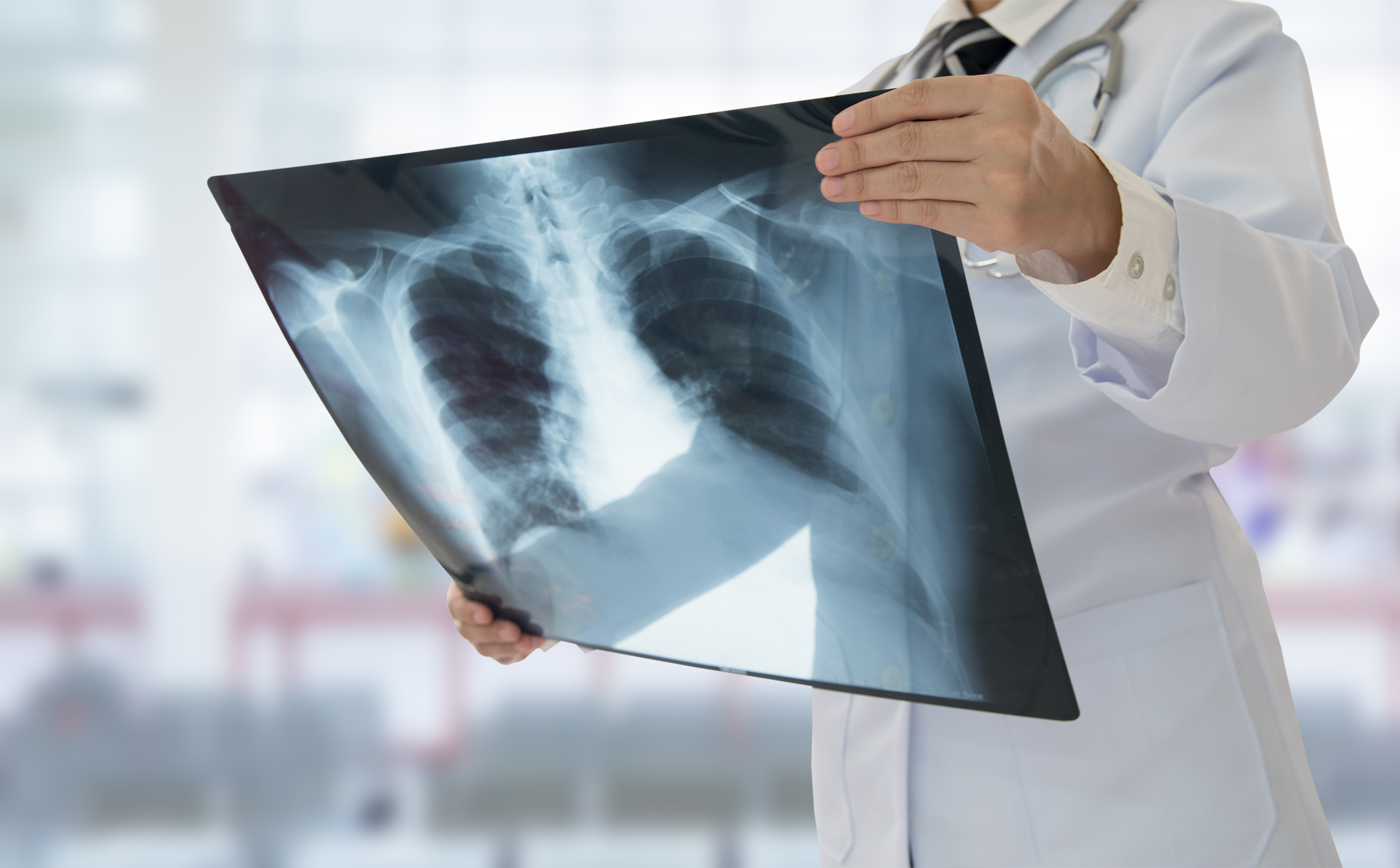
The Radiology department at Shaista Medical Center provides advanced diagnostic imaging services with rapid turnaround times. Our X-ray facilities are equipped with modern imaging equipment to produce high-quality images that support accurate diagnosis across all medical specialties.
We offer a comprehensive range of radiological services at affordable rates, ensuring that our patients receive timely and accurate diagnostic results. Our radiology team works closely with all clinical departments to provide integrated diagnostic support.
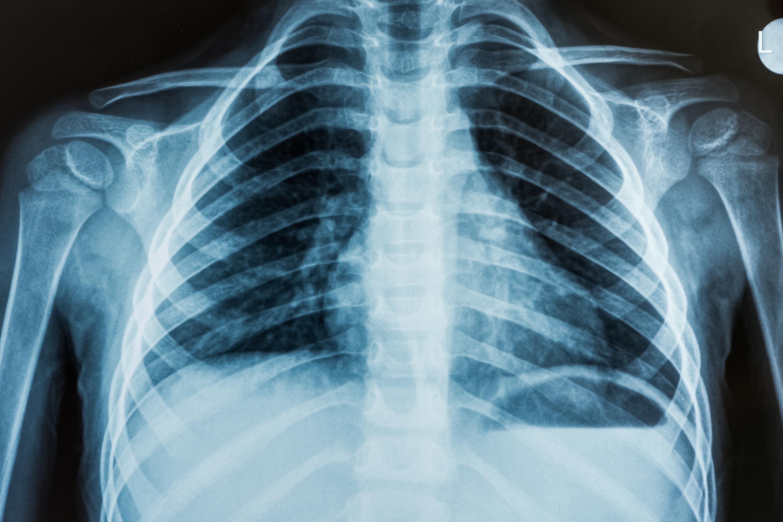
Chest X-Ray
Rapid chest X-ray imaging for respiratory conditions, pneumonia, and cardiac assessment.
Skeletal X-Rays
All bone and joint X-rays for fracture detection, arthritis, and orthopedic evaluation.
Abdominal X-Ray
Plain abdominal films for diagnosis of bowel obstruction, kidney stones, and other conditions.
Rapid Results
Quick image processing and reporting to minimize waiting time for patients and doctors.
X-Ray Services
Fast, affordable diagnostic imaging
Availability
X-Ray services available during hospital operating hours. Emergency imaging available 24/7.
Affordable Rates
Competitive pricing for all radiological procedures — quality care at affordable cost.
Department Gallery